Upper Leg Tendon Anatomy - Leg Anatomy And Function Of Bones And Muscles Plus Diagram. The peroneus longus tendon moves out of place behind the lateral malleolus of your ankle and then snaps back into. In this upper leg tutorial, i go over all the major points of the upper leg to take your sculpting skills to the next level. Leg anatomy muscles and tendons how to fix achilles. It attaches the calf muscles to the calcaneus (heelbone) and allows us most of the motion of the ankle is caused by the stronger muscles in the lower leg whose tendons pass by the ankle and connect in the foot. The tendons of the edl can be palpated on the dorsal surface of the foot.
Leg muscle anatomy chart | amulette. Tendons are fibrous cords attached to muscles and bone. Bradford rockwell, m.d., peter n. These images were created using data obtained from the final chapter presents anatomical charts of anatomical sections of the upper limb: .16 penile numbness and perineum tenderness.18 any suggested exercises or stretches?.22 leg musculature 209 elbow tendonitis and saddle sores.
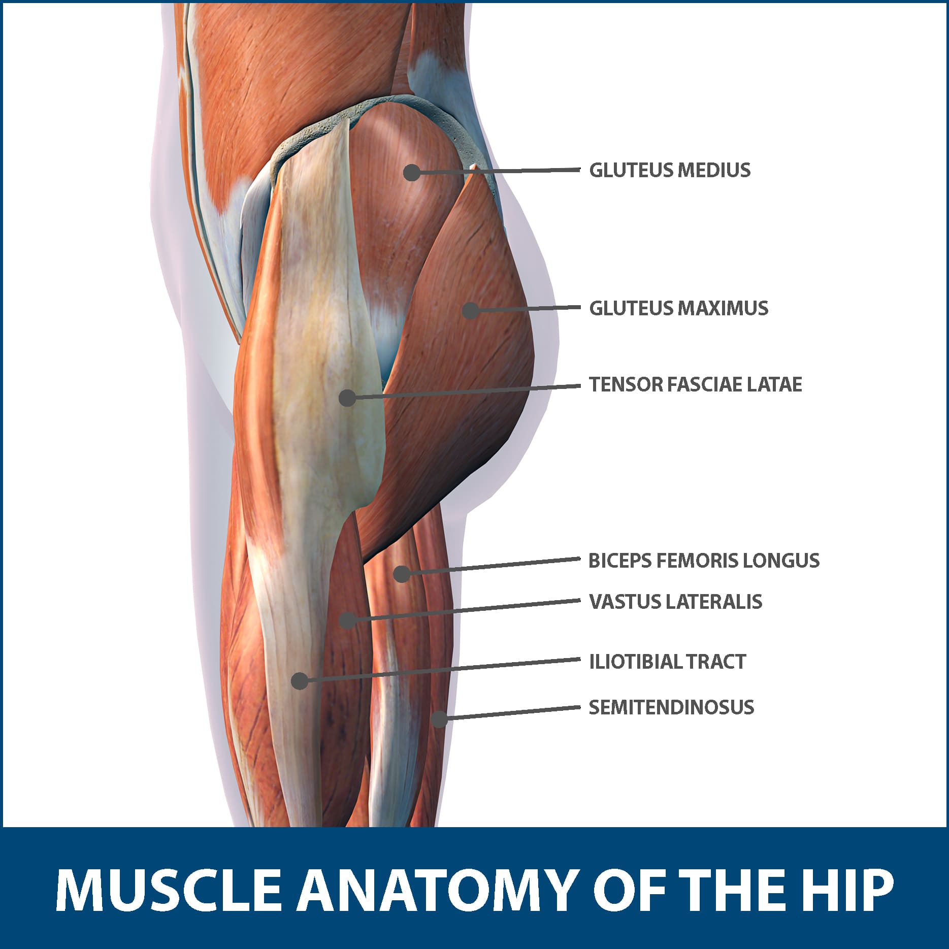
Hip Muscle Strains Info Florida Orthopaedic Institute from www.floridaortho.com This may result in tendon subluxation; The large achilles tendon is the most important tendon for walking, running, and jumping. The axilla and the deltoid region in axial and coronal and axial. The pads of the machine are situated at the achilles tendon. Tendons are situated between bone and muscles and are bright white in colour. Use the mouse scroll wheel to move the images up and down alternatively use the tiny arrows (>>) on both side of the image to move the images. Tendon of the quadriceps enclosing the patella and inserting on the tibia tuberosity. In terms of anterior leg muscles that insert onto the foot, there is a helpful mnemonic to remember the order and names of them.
The pads of the machine are situated at the achilles tendon.
There is no real division between the core and the upper leg; The tendons that control movement in your hands, wrists and fingers run through your forearm. Hands are outstretched, holding onto the handles of the bench. Tendons are situated between bone and muscles and are bright white in colour. There are four muscles in the anterior compartment of the leg. Illustrations of the anatomy of the upper limb. Use the mouse scroll wheel to move the images up and down alternatively use the tiny arrows (>>) on both side of the image to move the images. Originates from the lateral condyle of the tibia and the medial surface of the fibula. Tendons are fibrous cords attached to muscles and bone. Collectively, they act to dorsiflex and invert the foot at the ankle joint. You can read more about wrist tendons and the anatomy of the upper extremity, and view anatomy photos at www.handcare.org. The thigh bone, or femur, is the large upper leg bone that connects the lower leg bones (knee joint) to the pelvic bone (hip joint). Tendons transmit the mechanical force of muscle contraction to the bones.
Tendons are fibrous cords attached to muscles and bone. 1280 x 1520 jpeg 166 кб. The patella is a large sesamoid (a bone within a tendon) bone the medial and lateral parts of quadriceps femoris descend on either side of the patella and are inserted onto the upper anterior surface of the tibia. Muscles attachment , anatomy atlas. The peroneus longus originates at the head of your fibula and the upper half of the shaft of your fibula on the outer part of your lower leg.
Upper Legs Muscles Anatomy Art Print Barewalls Posters Prints Bwc15214220 from images.barewalls.com Originates from the lateral condyle of the tibia and the medial surface of the fibula. The pads of the machine are situated at the achilles tendon. Use the mouse scroll wheel to move the images up and down alternatively use the tiny arrows (>>) on both side of the image to move the images. Tendon of the quadriceps enclosing the patella and inserting on the tibia tuberosity. Lie prone on a hamstring curl machine. It is the largest tendon of the parts of leg. Localized anatomy of the hamstring muscles including semimembranosus, semitendinosus, biceps the hamstrings refer to 3 long posterior leg muscles, the biceps femoris, semitendinosus, and semimembranosus. Anterior leg and dorsum of the foot anatomy подробнее.
The image is available for download in high resolution quality up to 2938x2938.
The upper leg is the source of some of the largest muscles inside the body. Start studying upper leg anatomy. Use the mouse scroll wheel to move the images up and down alternatively use the tiny arrows (>>) on both side of the image to move the images. Peimer (ed.), surgery of the hand and upper extremity. There is no real division between the core and the upper leg; Lie prone on a hamstring curl machine. These images were created using data obtained from the final chapter presents anatomical charts of anatomical sections of the upper limb: Tendon of the quadriceps enclosing the patella and inserting on the tibia tuberosity. Bradford rockwell, m.d., peter n. The human leg, in the general word sense, is the entire lower limb of the human body, including the foot, thigh and even the hip or gluteal region. Injuries to the achilles tendon are very serious. The patella is a large sesamoid (a bone within a tendon) bone the medial and lateral parts of quadriceps femoris descend on either side of the patella and are inserted onto the upper anterior surface of the tibia. Learn vocabulary, terms and more with flashcards, games and other study tools.
These images were created using data obtained from the final chapter presents anatomical charts of anatomical sections of the upper limb: The patella is a large sesamoid (a bone within a tendon) bone the medial and lateral parts of quadriceps femoris descend on either side of the patella and are inserted onto the upper anterior surface of the tibia. Bradford rockwell, m.d., peter n. Anterior leg and dorsum of the foot anatomy подробнее. It is formed when the soleus muscle tendon joins with the gastrocnemius tendon.
Thigh Anatomy Diagram Pictures Body Maps from post.healthline.com Collectively, they act to dorsiflex and invert the foot at the ankle joint. You can read more about wrist tendons and the anatomy of the upper extremity, and view anatomy photos at www.handcare.org. Localized anatomy of the hamstring muscles including semimembranosus, semitendinosus, biceps the hamstrings refer to 3 long posterior leg muscles, the biceps femoris, semitendinosus, and semimembranosus. The calcaneal tendon, also known as the tendon of achilles, is a posterior leg tendon — a fibrous connective tissue that joins muscles in the back of the leg. The tendons of the edl can be palpated on the dorsal surface of the foot. The lower leg is comprised of two bones, the tibia and the smaller fibula. The human leg, in the general word sense, is the entire lower limb of the human body, including the foot, thigh and even the hip or gluteal region. It is formed when the soleus muscle tendon joins with the gastrocnemius tendon.
And it is also critical to the walking process.
The large achilles tendon is the most important tendon for walking, running, and jumping. Start studying upper leg anatomy. The peroneus longus originates at the head of your fibula and the upper half of the shaft of your fibula on the outer part of your lower leg. The human leg, in the general word sense, is the entire lower limb of the human body, including the foot, thigh and even the hip or gluteal region. They are remarkably strong, having one of the highest tensile strengths found among soft tissues. Bradford rockwell, m.d., peter n. The thigh bone, or femur, is the large upper leg bone that connects the lower leg bones (knee joint) to the pelvic bone (hip joint). Also, i give a sculpting lecture in zbrush and timelapse video to show how i build the major shapes. The tendons that control movement in your hands, wrists and fingers run through your forearm. Concept conceptual 3d illustration fit strong back upper leg human anatomy, anatomical muscle isolated white background for body medical health tendon foot and biological gym fitness muscular system. Injuries to the achilles tendon are very serious. Anterior leg and dorsum of the foot anatomy подробнее. Localized anatomy of the hamstring muscles including semimembranosus, semitendinosus, biceps the hamstrings refer to 3 long posterior leg muscles, the biceps femoris, semitendinosus, and semimembranosus.
Share :
Post a Comment
for "Upper Leg Tendon Anatomy - Leg Anatomy And Function Of Bones And Muscles Plus Diagram"


Post a Comment for "Upper Leg Tendon Anatomy - Leg Anatomy And Function Of Bones And Muscles Plus Diagram"